Glaucoma Monitoring and Testing

Senior woman with glasses thinking

Monitoring Glaucoma: The key to preventing further vision loss

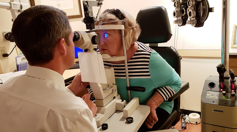
Carefully monitoring the progression of your glaucoma is a significant step in preventing vision loss. Subtle changes that occur in the eye which may not be noticeable to you, must be detected promptly to prevent vision loss. As a patient of the Glaucoma Program at Tyson Eye, you may be asked to return to see your doctor every three months, depending on your stage of glaucoma. At these visits, different diagnostic tests will be performed and compared to the results of your previous tests. These tests are done using state-of-the-art equipment, most of which is computer assisted and capable of providing greatly detailed information about the health of your eyes and your optic nerves. The Tyson Eye Glaucoma Program is a recommended plan to monitor the progression of glaucoma. It is similar to the programs at most major university eye departments.

As a glaucoma patient at Tyson Eye, you are one of many patients that are part of a structured Glaucoma Program in Southwest Florida. We understand the importance of preserving your precious vision and want to do everything in our power to help you escape the damage caused by “the silent thief of sight,” also known as glaucoma.

What you can do to protect your sight from glaucoma:

  • Follow the recommended pressure check regimen prescribed by your doctor. Be sure to schedule your appointments and return to the office for your scheduled pressure checks and eye examinations.
  • Use your medications as directed at all times. Stay informed and learn as much as you can about glaucoma.
  • Call the Glaucoma Program Team at Tyson Eye if you have any questions or concerns.
A woman having her eyes examined by an optometrist

Goals of our Glaucoma Program

  • To provide a customized treatment plan to monitor the progression of your glaucoma.
  • To provide state-of-the-art technology while maintaining the old-fashioned concern for which Tyson Eye is recognized.
  • To deliver to all of our patients long-term maximum preservation of vision. We understand your eye-sight is precious.
  • To provide a specialized team of ophthalmic professionals dedicated to monitoring your compliance.
  • To create accessibility and familiarity with our staff so that you have answers to all of your questions.

The Tyson Eye Glaucoma Program Schedule

(Based on programs at major university eye departments)

Quarter 1

Comprehensive Eye Examination (including refraction)

Fundus Photos

Quarter 2

Pressure Check

Visual Field Exam Gonioscopy

Quarter 3

Pressure Check

Ocular Blood Flow Analysis

Quarter 4

Pressure Check

GDX or OCT

Treatment Options for Glaucoma

Medications

Eye drops can be used effectively to help maintain and keep your eye at a healthy eye pressure and are an important part of the treatment routine for most people with glaucoma. A variety of glaucoma eye drops are now available by prescription that either increase the drainage of the aqueous humor or slow its production. Sometimes a combination of eye drops is prescribed to obtain the best results.

Laser Treatments

Various lasers are available to treat the different types of glaucoma. In the United States, laser treatment is growing in popularity as the primary treatment for glaucoma, even over eye drop medications. In many cases, it reduces or eliminates the need for daily drops, a necessary daily ritual that many find expensive and undesirable.

  • Selective Laser Trabeculoplasty (SLT)
    One of the newest advances in treating open angle glaucoma. Similar to the ALT, this laser works on the trabecular meshwork to improve drainage. It selects only certain cells to minimize damage to surrounding cells and can be repeated several times if needed. It is appropriate for patients who have or have not had their cataracts removed.
  • Laser Peripheral Iridotomy (LPI)
    This type of laser surgery is used to treat narrow angle glaucoma. This laser treatment involves making a tiny hole in the iris to allow improved drainage of the aqueous humor.

Minimally Invasive Glaucoma Surgery (MIGS)

Dr. Tyson and Dr. Stephens specialize in the treatment of glaucoma through minimally invasive glaucoma surgery (MIGS) which are designed to manage glaucoma in a safer and more natural way than other glaucoma treatments. MIGS treatments can also reduce the dependence of medicated eye drops, benefiting the patient by saving money over time and having fewer daily interruptions to apply eye drops. There are a number of MIGs devices that your surgeon can choose from depending on the characteristics of your eyes.

  • iStent, is the first device that improves your eye’s natural fluid outflow to safely lower eye pressure by creating a permanent opening in the eye meshwork.
  • Hydrus, expands the eye’s natural drainage canal.
  • Kahook, a goniotomy blade created to produce a more complete removal of trabecular meshwork through a minimally invasive approach.
  • Xen, a surgical implant designed to lower high eye pressure in open angle glaucoma patients.
  • Omni, is designed to perform two implant free procedures during a single procedure. Canaloplasty of Schlemm’s canal followed by trabeculotomy of the trabecular meshwork to reduce intraocular pressure in adult patients with primary open angle glaucoma.
  • ECP, the ciliary body of the eye, which creates fluid, is treated with a laser.

Traditional Surgeries

Seldom used today due to new advances in laser treatment for glaucoma, these traditional surgical procedures are usually turned to as a last resort when all other treatment methods have failed to slow the progression of the glaucoma. These surgical procedures must be done in an operating room under sterile conditions to minimize the risk of infection and complications.

  • Trabeculectomy
    Trabeculectomy or “filtering surgery,” involves removing a piece of tissue from the sclera (the white part of your eye). This creates a “flap valve” in the trabecular meshwork. This new pathway allows fluid to flow out more easily. This fluid pools in a little bubble under the eyelid, called a “bleb.” There, it is reabsorbed into the body via the circulatory system. Your surgeon may elect to insert a tube shunt to prevent the bleb from closing.
  • Tube shunt
    A flexible glaucoma drainage device that is implanted in the eye to regulate the fluid pressure inside of the eye.